Introducing

R-FIX

Rib-Based Spinal Fixation

A fundamentally new approach to spinal deformity correction that utilizes the thoracic cage as an active load-sharing component.

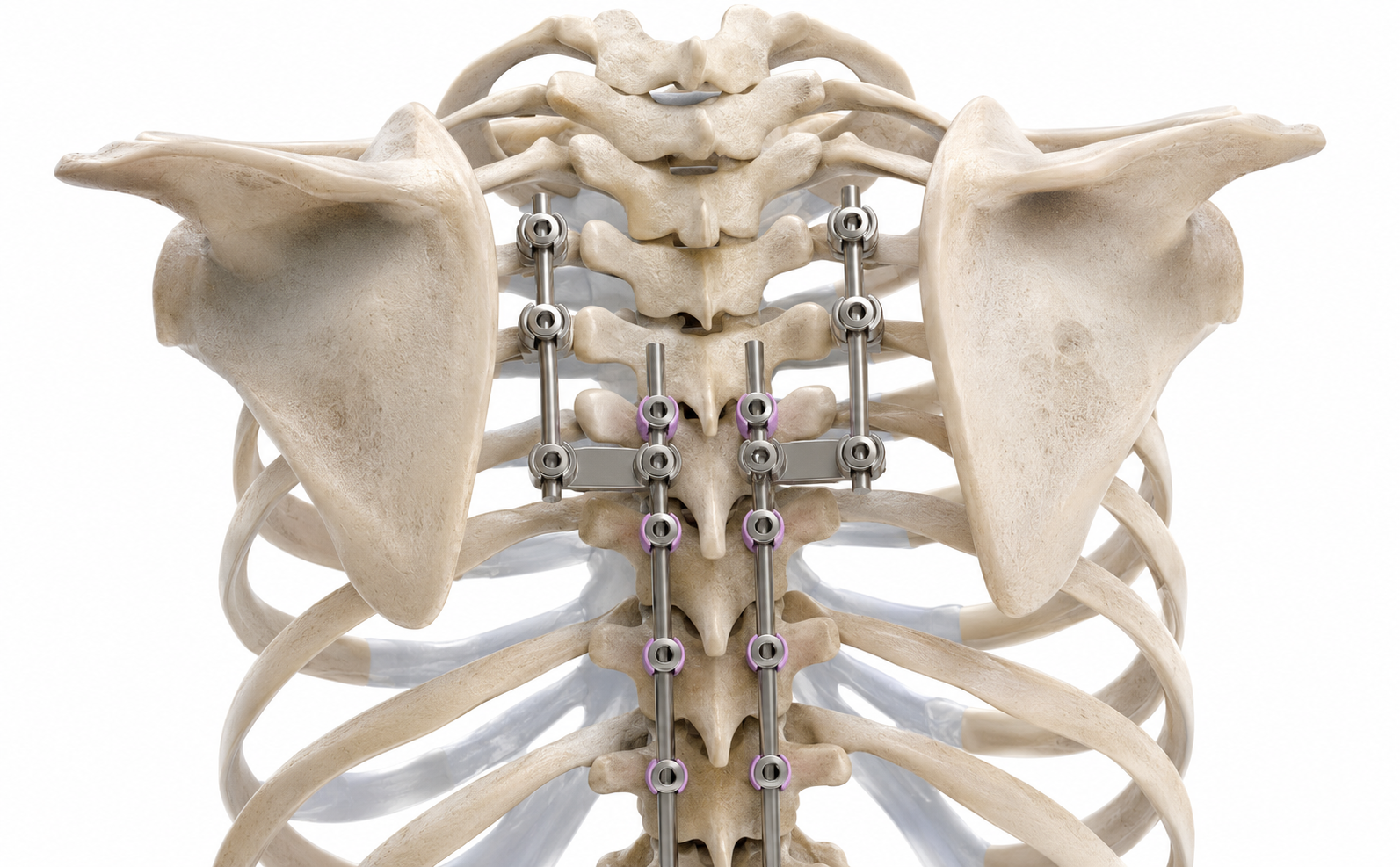
R-FIX construct engaging the thoracic spine and rib cage

Clinical Challenges

Engineered to address two of the most challenging problems in spinal deformity surgery.

Pediatric

Early-Onset Spinal Deformity

Immature pedicles, growth-preservation, and long-construct instability create challenges that conventional fixation was not designed to address.

Explore EOS

Adult

Adult Spinal Deformity & PJK Prevention

Long thoracolumbar constructs concentrate load at the upper instrumented vertebra, contributing to proximal junctional failure.

Explore Adult Applications

Biomechanical Rationale

Why Engage the Thoracic Cage?

Conventional pedicle-based fixation and rib-based fixation each provide unique biomechanical characteristics. Rib-based fixation utilizes the structural strength of the thoracic cage to supplement spinal fixation, redistribute forces, and address limitations of conventional pedicle-only constructs in selected pediatric and adult deformity patients.

Early-Onset Spinal Deformity

Pediatric Deformity Comparison

Conventional

Pedicle-Based Fixation

Radiograph — Pedicle Screw Pullout
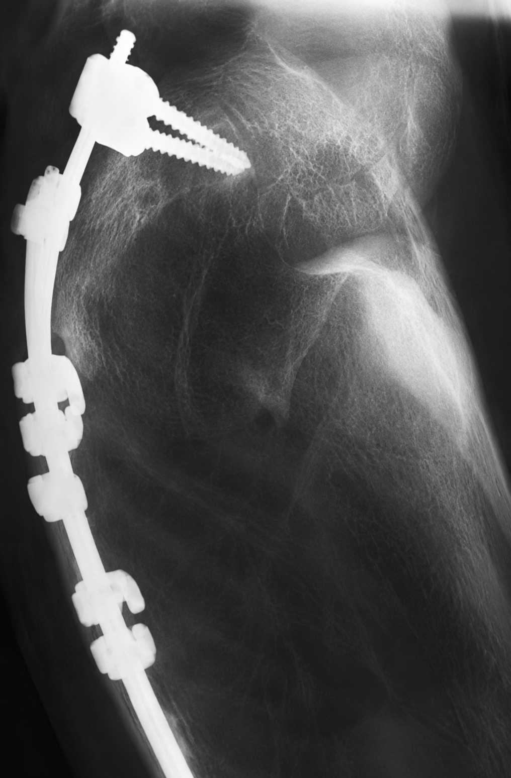
Pedicle Screw Pullout. Screw loosening and pullout at proximal anchors in an immature spine.
Radiograph — Rod Fracture
Rod Fracture. Rod failure adjacent to a rigid pedicle-only construct.
Radiograph — Neurological Injury
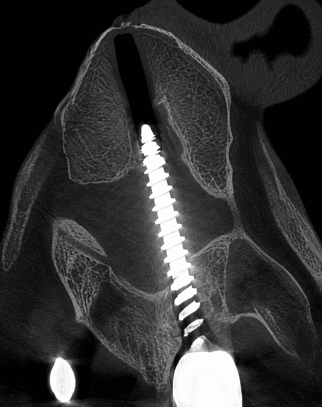
Neurological Injury. Malpositioned pedicle screw with breach into the spinal canal.

Representative Challenges

  • High construct stiffness
  • Increased autofusion risk
  • Pedicle screw pullout
  • Rod fracture
  • Proximal junctional kyphosis
  • Immature pedicles
  • Neurological injury risk

Rib-Based Fixation

Thoracic Cage Engagement

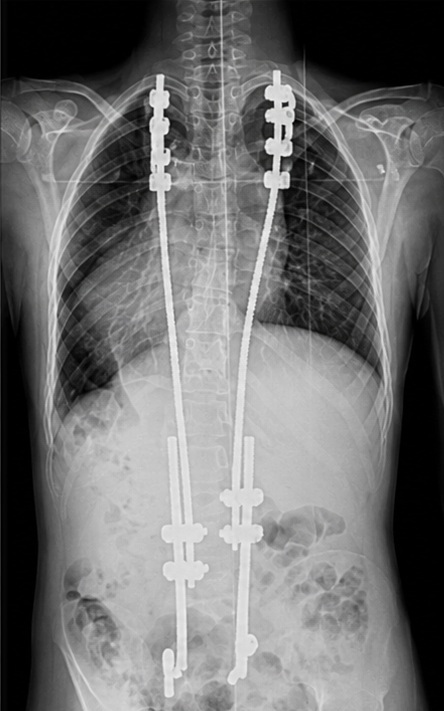
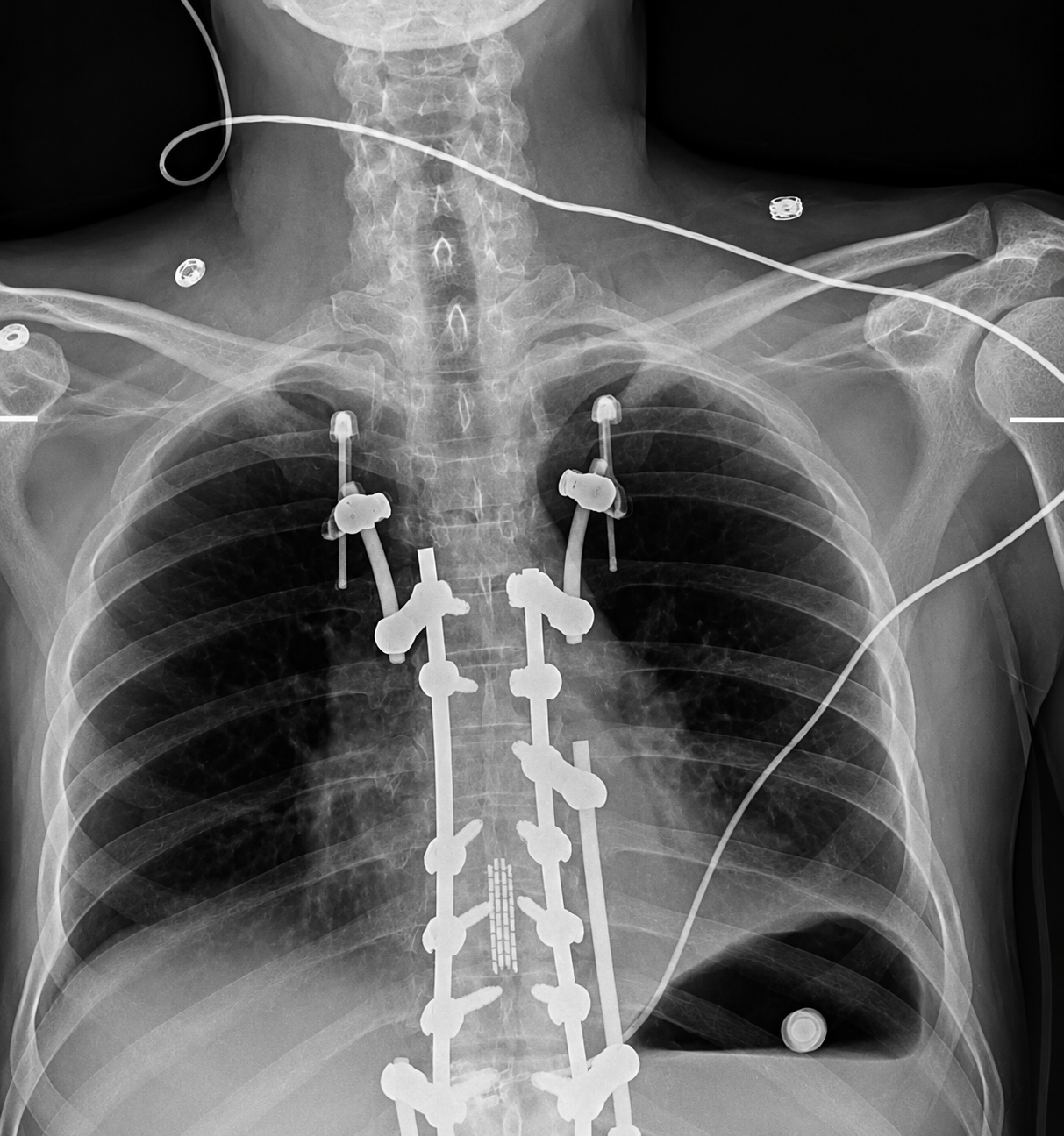
Radiograph — Rib-Based Fixation
Rib-Based Fixation. AP radiograph showing bilateral rib-based spinal fixation rods.
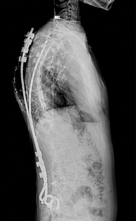
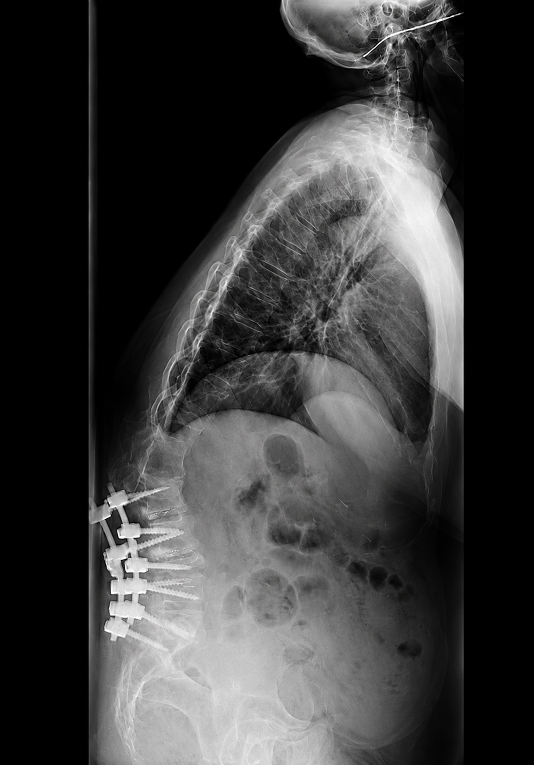
Radiograph — Rib-Based Fixation
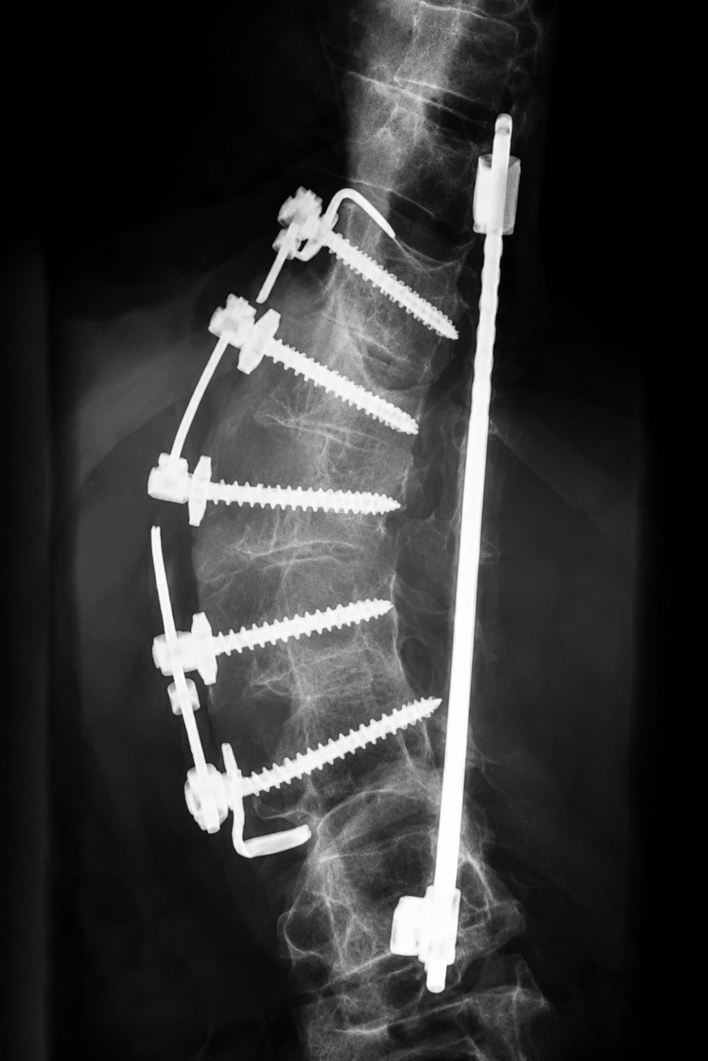
Rib-Based Fixation. Lateral radiograph showing bilateral rib-based spinal fixation rods.

Biomechanical Advantages

  • Lower construct stiffness
  • Growth-friendly fixation
  • Increased pullout resistance
  • Reduced rod fracture risk
  • Reduced proximal junctional kyphosis
  • Avoids immature pedicles
  • Reduced neurological risk

Adult Spinal Deformity

Adult Deformity Comparison

Conventional

Pedicle-Based Fixation

Radiograph — Proximal Junctional Kyphosis
Proximal Junctional Kyphosis. PJK developing above a long thoracolumbar construct.
Radiograph — Pedicle Screw Pullout
Pedicle Screw Pullout. Anchor pullout at the UIV under transition-zone stress.
Radiograph — Proximal Junctional Failure
Proximal Junctional Failure. Structural failure at the upper instrumented vertebra.

Representative Challenges

  • Abrupt construct transition
  • Stress concentration at the UIV
  • Damage to posterior ligamentous complex
  • Facet joint disruption
  • Paraspinal muscle injury
  • Pedicle screw pullout
  • High PJK/PJF risk

Published studies have reported proximal junctional kyphosis rates approaching 50% in selected high-risk adult spinal deformity populations, depending on patient characteristics, construct design, and clinical definitions.

Rib-Based Fixation
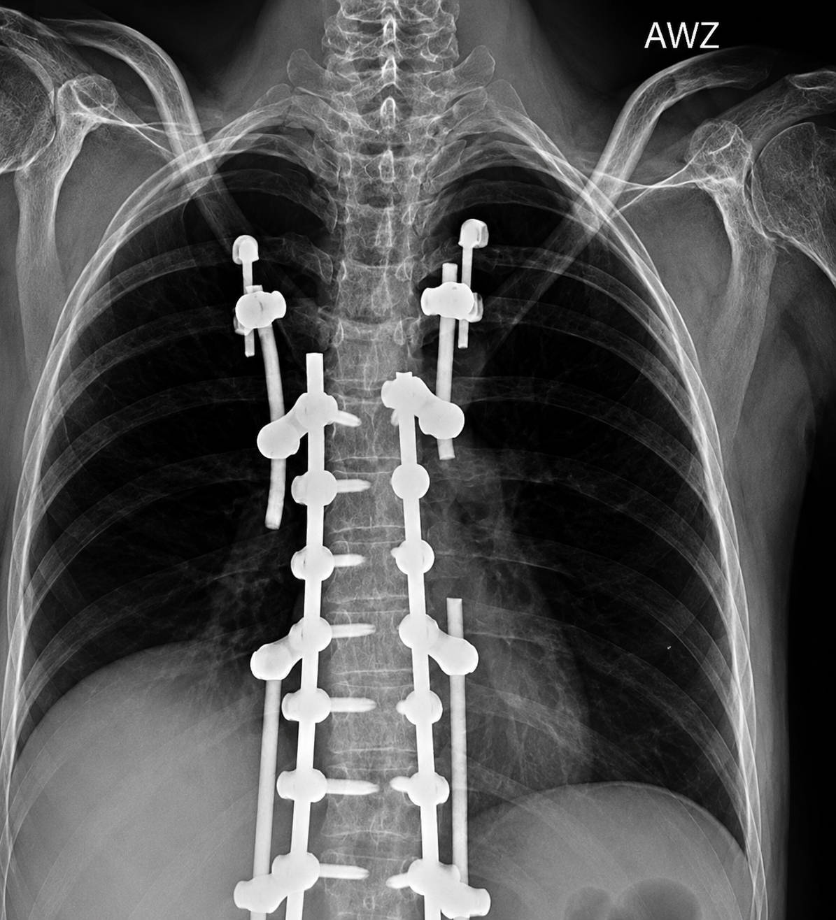
Thoracic Cage Engagement

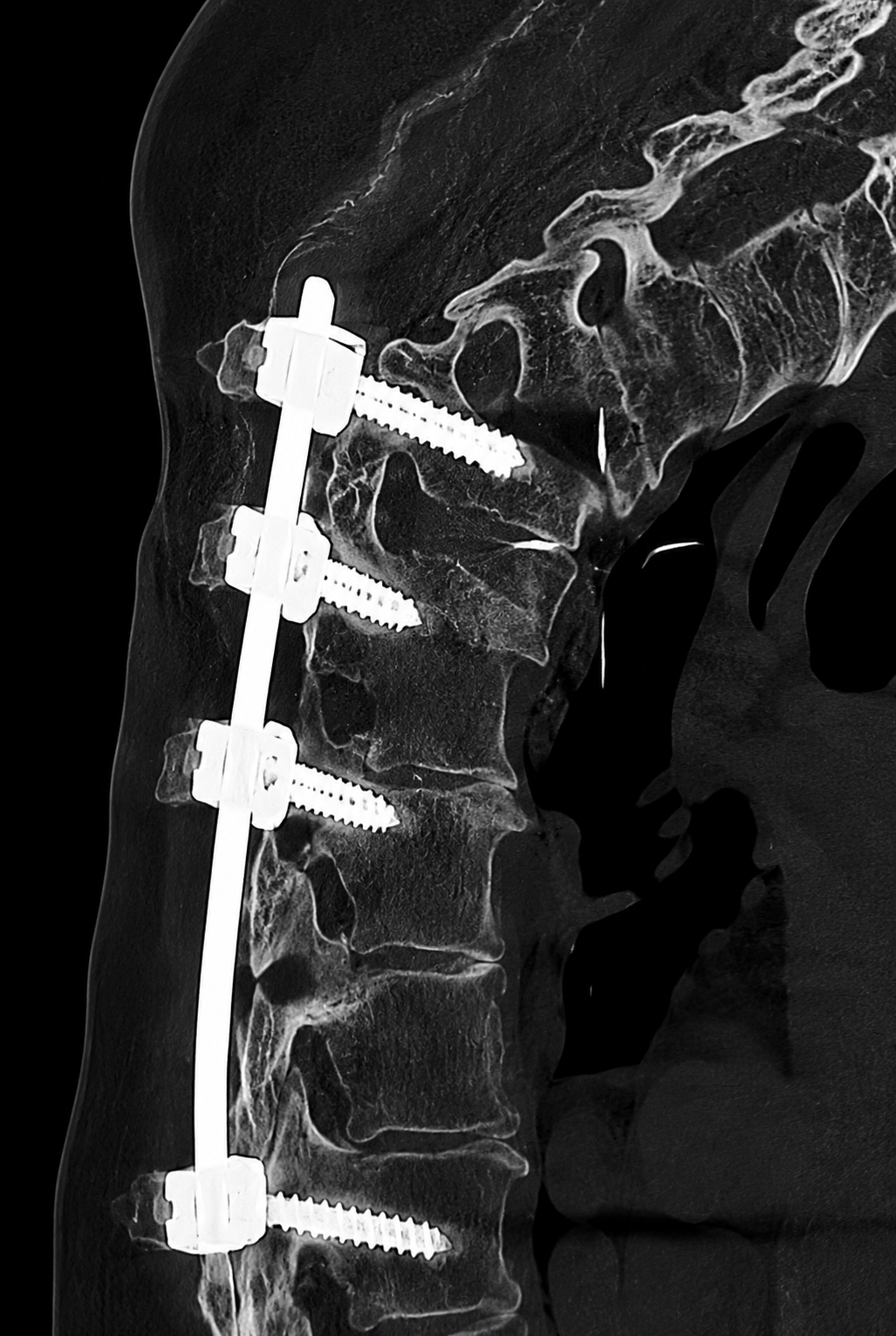
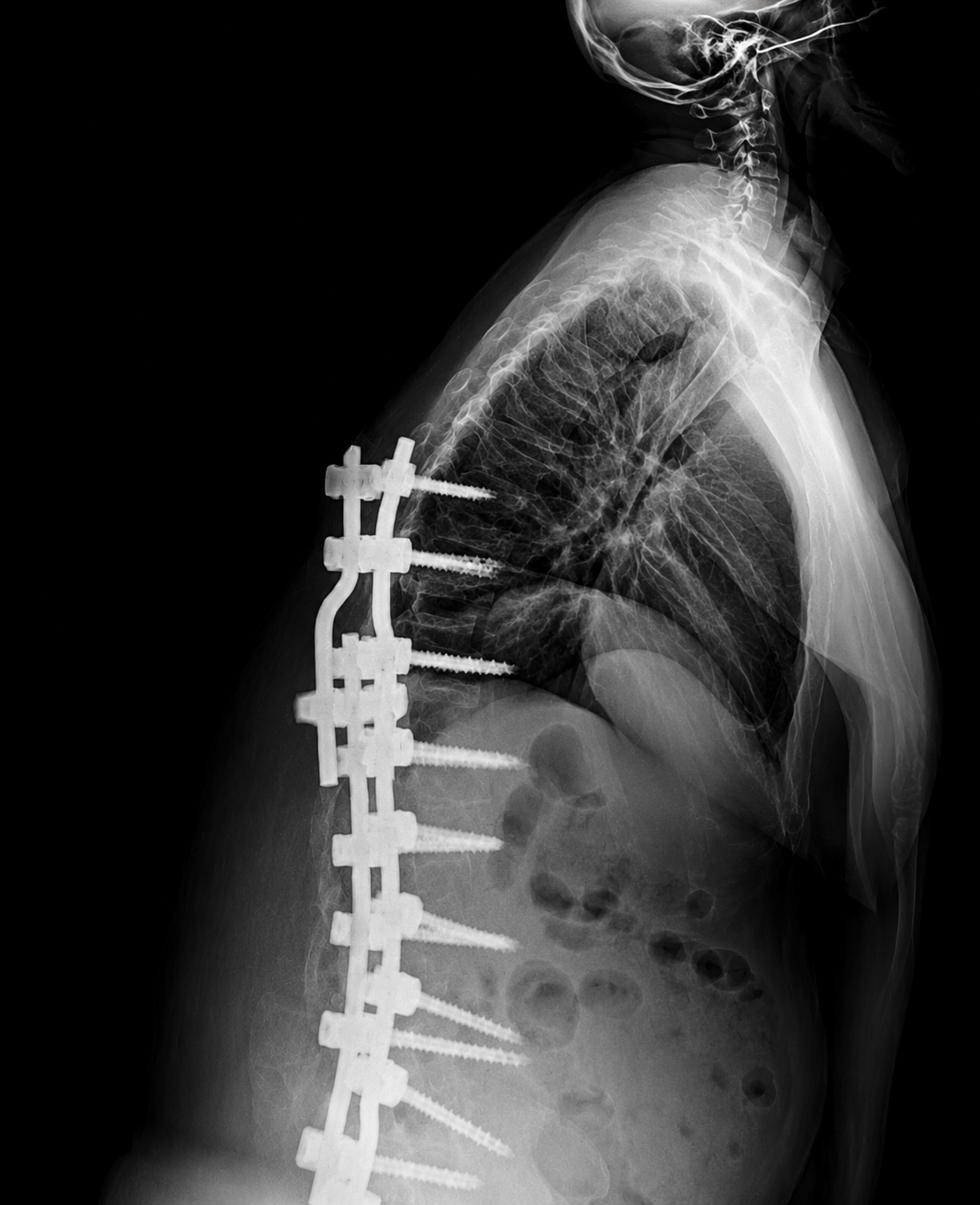
Radiograph — Rib-Based Fixation
Rib-Based Fixation. Long thoracolumbar construct with rib-based upper anchors.
Radiograph — Rib-Based Fixation
Rib-Based Fixation. Rib-based UIV anchoring for PJK prevention.

Biomechanical Advantages

  • Establishes a "soft landing"
  • Reduces stress concentration
  • Limits damage to posterior ligamentous complex
  • Eliminates UIV facet joint disruption
  • Preserves paraspinal musculature
  • Increased pullout resistance
  • Designed to address biomechanical factors associated with PJK/PJF

Engineering Principles

The thoracic cage can function as an active component of the fixation construct rather than simply surrounding the spine.

Broader Load Sharing

Distribute construct forces across multiple rib attachment points to reduce peak stress at the bone–implant interface.

Gradual Transition Zone

Soften the mechanical transition between instrumented and non-instrumented segments to help mitigate junctional failure.

Extra-Spinal Fixation

Engage anatomy outside the vertebral column to preserve spinal motion and biologic potential where appropriate.

Purpose-Built Design

Engineered around rib anatomy.

Unlike traditional spinal hooks, which were adapted for rib fixation but were not specifically engineered around rib anatomy, R-FIX™ is the first anatomic rib-anchor platform engineered specifically for the correction and stabilization of spinal deformity. Every implant and instrument was designed to improve anatomical conformity, load distribution, and surgical versatility across pediatric and adult applications.
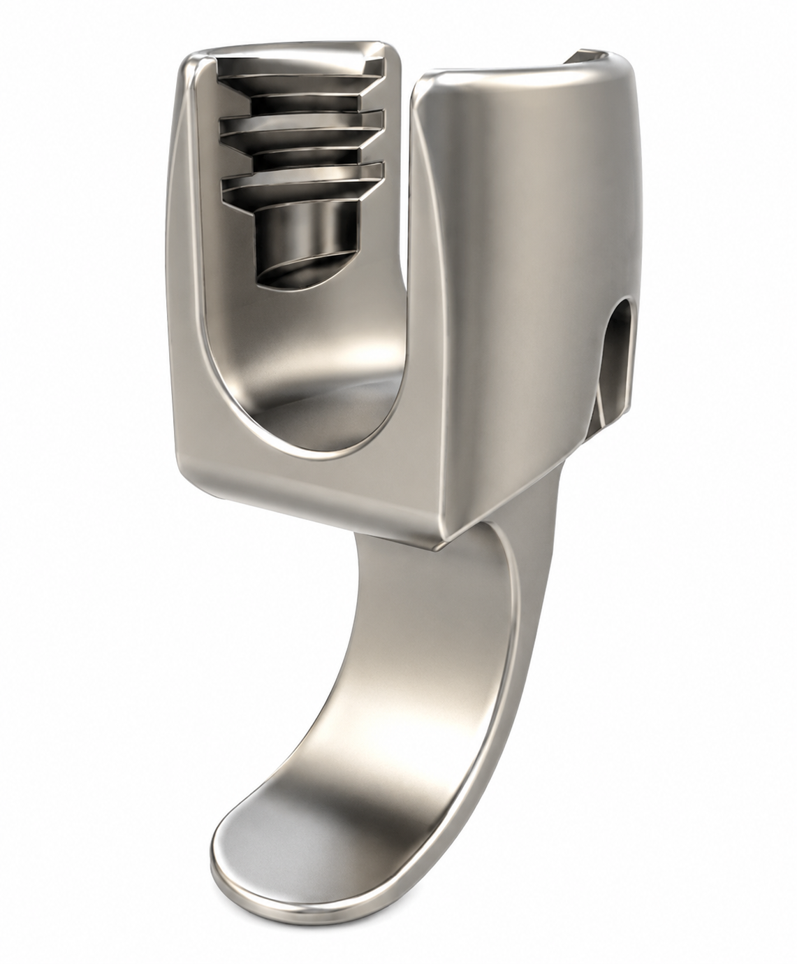
Traditional spinal hook, originally designed for spinal fixation and later adapted for rib fixation

Traditional Spinal Hook

Originally designed for spinal fixation and later adapted for rib fixation, with limited conformity to native rib anatomy.
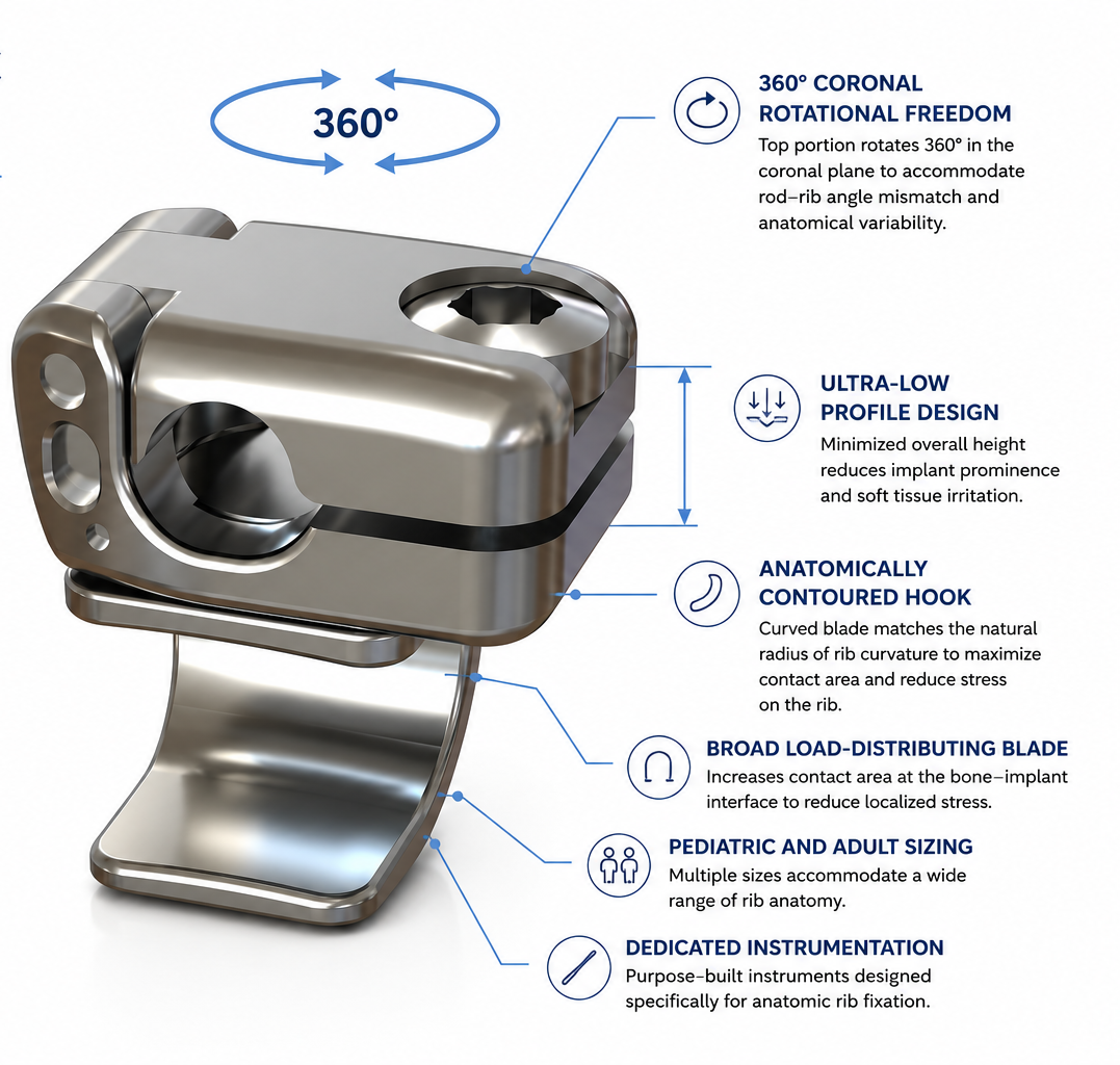
R-FIX anatomic rib anchor with engineering callouts: 360° coronal rotational freedom, ultra-low profile, anatomically contoured anchor, broad load-distributing blade, pediatric and adult sizing, dedicated instrumentation

R-FIX™ Anatomic Rib Anchor

Purpose-designed around rib anatomy for spinal deformity correction.

Technology Platform

The R-FIX platform.

A comprehensive system of anatomic anchors, connectors, and instruments — engineered as one platform.

Swivel Anchor

Multi-axial rib engagement designed for anatomical variability.

Ultra-Low Profile Anchor

Reduced prominence for thin soft-tissue envelopes.

Uniaxial Anchor

Single-plane rib anchor providing predictable engagement with a streamlined footprint.

Monoaxial Anchor

Fixed-angle interface for controlled construct geometry.

Monoaxial Extra Wide Anchor

Extra-wide fixed-angle rib anchor engineered for enhanced load distribution across broader rib anatomy.

Connectors

Modular linkage between rib anchors and the primary construct.

Sliding Connector

Accommodates the spectrum of anatomic distance between rib anchors and primary construct.

Pedicle Screw System

Integrated thoracolumbar anchor set compatible with R-FIX.

Instrumentation

Dedicated instrumentation designed specifically for anatomic rib fixation.

Clinical Applications
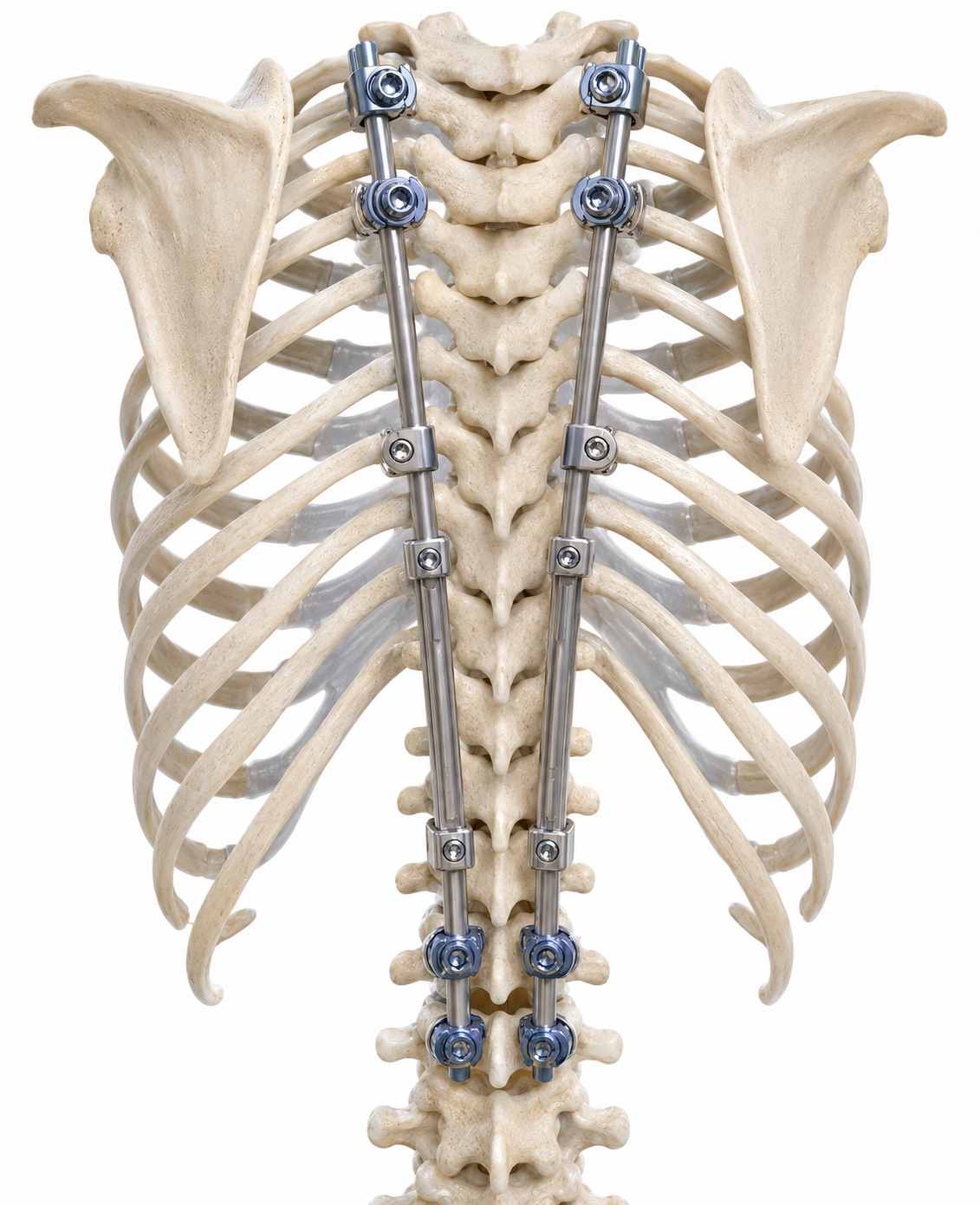
One platform. Two distinct clinical arenas.

EOS construct rendering — growing rod / hybrid configuration on pediatric thoracic spine

Pediatric

Early-Onset Spinal Deformity

  • Growing Rod Construct
  • Hybrid Construct
  • Growth-Friendly Fixation
  • Reduced Pedicle Dependence
  • Reduced Pedicle Screw Pullout, Rod Fracture, PJK, and Neurological Injury Risk

Adult

Adult Spinal Deformity & PJK Prevention

  • Soft Landing above UIV
  • Preserved Soft-Tissues and Posterior Tension Band
  • Load Sharing
  • Long Thoracolumbar Constructs
  • PJK Prevention
Adult construct rendering — long thoracolumbar with rib-based UIV for PJK prevention

Scientific Validation

Evidence at every stage.

  1. 01
  2. 02
  3. 03
  4. 04
  5. 05
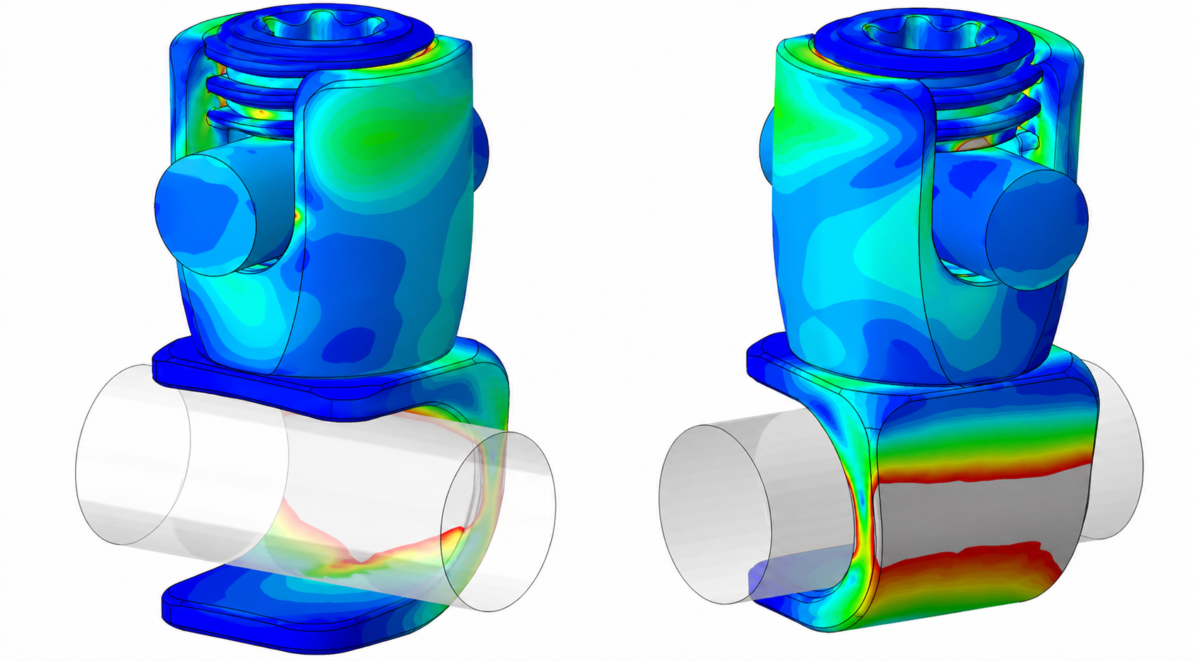
Finite Element Analysis — figure

Stage 01

Finite Element Analysis

Computational modeling of construct behavior across anatomical variations and loading conditions.

Select a stage above
Learn More

Clinical Evidence

A growing body of preclinical and clinical evidence.